

What Was The First Animal That Had A Brain?
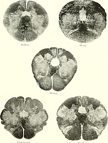
Evolution of the encephalon from ape to man
There is so much to be discovered about the evolution of the encephalon and the principles that govern information technology. While much has been discovered, not everything currently known is well understood. The evolution of the brain has appeared to exhibit diverging adaptations inside taxonomic classes such equally mammalia and more vastly diverse adaptations beyond other taxonomic classes. Brain to body size scales allometrically.[1] This means as torso size changes, so do other physiological, anatomical, and biochemical constructs connecting the encephalon to the body.[2] Small bodied mammals have relatively large brains compared to their bodies whereas big mammals (such as whales) accept smaller brain to trunk ratios. If brain weight is plotted confronting trunk weight for primates, the regression line of the sample points can indicate the encephalon ability of a primate species. Lemurs for example fall beneath this line which ways that for a primate of equivalent size, we would wait a larger brain size. Humans lie well above the line indicating that humans are more encephalized than lemurs. In fact, humans are more encephalized compared to all other primates.[three] This ways that human brains have exhibited a larger evolutionary increase in it complexity relative to its size. Some of these evolutionary changes have been found to be linked to multiple genetic factors such as, proteins and other organelles.
Early history of brain development [edit]
1 approach to understanding overall brain development is to use a paleoarchaeological timeline to trace the necessity for always increasing complication in structures that allow for chemical and electrical signaling. Because brains and other soft tissues practice not fossilize equally readily equally mineralized tissues, scientists frequently look to other structures as evidence in the fossil record to go an understanding of brain evolution. This, however, leads to a dilemma as the emergence of organisms with more than complex nervous systems with protective bone or other protective tissues that can so readily fossilize occur in the fossil record before prove for chemical and electrical signaling.[4] [v] Testify from 2008 showed that the ability to transmit electrical and chemical signals existed even before more than complex multicellular lifeforms.[four]
Fossilization of encephalon, or other soft tissue, is possible all the same, and scientists tin can infer that the get-go brain structure appeared at to the lowest degree 521 meg years ago, with fossil brain tissue nowadays in sites of infrequent preservation.[six]
Another approach to understanding brain evolution is to look at extant organisms that do not possess complex nervous systems, comparing anatomical features that allow for chemic or electrical messaging. For case, choanoflagellates are organisms that possess various membrane channels that are crucial to electrical signaling. The membrane channels of choanoflagellates' are homologous to the ones found in brute cells, and this is supported by the evolutionary connection between early choanoflagellates and the ancestors of animals.[4] Some other example of extant organisms with the chapters to transmit electric signals would be the glass sponge, a multicellular organism, which is capable of propagating electrical impulses without the presence of a nervous organisation.[7]
Before the evolutionary evolution of the brain, nerve nets, the simplest form of a nervous system developed. These nerve nets were a sort of precursor for the more evolutionarily avant-garde brains. They were showtime observed in Cnidaria and consist of a number of neurons spread apart that let the organism to answer to physical contact. They are able to rudimentarily find food and other chemicals merely these nerve nets do not allow them to detect the source of the stimulus.
Ctenophores also demonstrate this crude precursor to a brain or centralized nervous organization, however, they phylogenetically diverged before the phylum Porifera and Cnidaria. There are two current theories on the emergence of nervus nets. One theory is that nerve nets may have developed independently in Ctenophores and Cnidarians. The other theory states that a common ancestor may take adult nervus nets, but they were lost in Porifera. While comparison the average neuron size and the packing density the difference between primate and mammal brains is shown.[viii]
A tendency in encephalon evolution according to a written report done with mice, chickens, monkeys and apes ended that more evolved species tend to preserve the structures responsible for bones behaviors. A long term human study comparison the human brain to the primitive brain establish that the modern human being brain contains the primitive hindbrain region – what virtually neuroscientists call the protoreptilian brain. The purpose of this part of the brain is to sustain central homeostatic functions, which are cocky regulating processes organisms use to help their bodies conform. The pons and medulla are major structures establish at that place. A new region of the encephalon developed in mammals nearly 250 million years subsequently the appearance of the hindbrain. This region is known as the paleomammalian brain, the major parts of which are the hippocampi and amygdalas, often referred to as the limbic arrangement. The limbic arrangement deals with more complex functions including emotional, sexual and fighting behaviors. Of form, animals that are not vertebrates also take brains, and their brains accept undergone split up evolutionary histories.[six]
The brainstem and limbic system are largely based on nuclei, which are essentially balled-up clusters of tightly packed neurons and the axon fibers that connect them to each other, equally well as to neurons in other locations. The other two major brain areas (the cerebrum and cerebellum) are based on a cortical architecture. At the outer periphery of the cortex, the neurons are bundled into layers (the number of which vary according to species and office) a few millimeters thick. There are axons that travel between the layers, but the majority of axon mass is below the neurons themselves. Since cortical neurons and most of their axon fiber tracts don't have to compete for space, cortical structures tin can scale more hands than nuclear ones. A key feature of cortex is that because it scales with surface area, more of it can be fit inside a skull by introducing convolutions, in much the same way that a dinner napkin can be blimp into a glass by wadding information technology upwards. The degree of convolution is by and large greater in species with more complex behavior, which benefits from the increased surface area.
The cerebellum, or "little brain," is backside the brainstem and below the occipital lobe of the cerebrum in humans. Its purposes include the coordination of fine sensorimotor tasks, and it may be involved in some cognitive functions, such every bit language and different motor skills that may involve hands and feet. The cerebellum helps proceed equilibrium. Damage to the cerebellum would consequence in all physical roles in life to be affected. Homo cerebellar cortex is finely convoluted, much more so than cognitive cortex. Its interior axon cobweb tracts are called the arbor vitae, or Tree of Life.
The area of the brain with the greatest amount of recent evolutionary modify is called the neocortex. In reptiles and fish, this area is chosen the pallium, and is smaller and simpler relative to trunk mass than what is found in mammals. According to research, the cerebrum offset developed about 200 million years agone. It's responsible for college cognitive functions - for example, language, thinking, and related forms of information processing.[nine] It'southward also responsible for processing sensory input (together with the thalamus, a part of the limbic system that acts as an information router). The thalamus receives the different sensations before it is then passed onto the cerebral cortex. Most of its role is subconscious, that is, not available for inspection or intervention by the conscious mind. The neocortex is an elaboration, or outgrowth, of structures in the limbic system, with which it is tightly integrated. The neocortex is the main part controlling many brain functions as it covers half of the whole encephalon in volume. The development of these recent evolutionary changes in the neocortex were likely developed as a result of new neural network formations and positive selections of certain genetic components.
Role of embryology in the development of the brain [edit]
In addition to studying the fossil record, evolutionary history can exist investigated via embryology. An embryo is an unborn/unhatched animal and evolutionary history can be studied by observing how processes in embryonic evolution are conserved (or not conserved) beyond species. Similarities between different species may indicate evolutionary connexion. One mode anthropologists study evolutionary connection betwixt species is by observing orthologs. An ortholog is defined as two or more homologous genes betwixt species that are evolutionarily related by linear descent. By using embryology the evolution of the encephalon tin be tracked between various species.
Os morphogenetic poly peptide (BMP), a growth factor that plays a significant function in embryonic neural development, is highly conserved amongst vertebrates, every bit is sonic hedgehog (SHH), a morphogen that inhibits BMP to allow neural crest development. Tracking these growth factors with the use of embryology provides a deeper understanding of what areas of the brain diverged in their evolution. Varying levels of these growth factors atomic number 82 to differing embryonic neural development which and so in plough affects the complication of hereafter neural systems. Studying the brain'due south development at various embryonic stages across differing species provides additional insight into what evolutionary changes may have historically occurred. This and then allows scientists to look into what factors may take caused such changes, such every bit links to neural network diversity, growth gene production, protein- coding selections, and other genetic factors.
Randomizing admission and scaling brains upward [edit]
Some animate being phyla accept gone through major encephalon enlargement through evolution (e.g. vertebrates and cephalopods both contain many lineages in which brains have grown through development) merely most animal groups are composed only of species with extremely modest brains. Some scientists[ who? ] argue that this deviation is due to vertebrate and cephalopod neurons having evolved means of communicating that overcome the scalability problem of neural networks while well-nigh animal groups have not. They argue that the reason why traditional neural networks fail to improve their office when they scale up is because filtering based on previously known probabilities cause cocky-fulfilling prophecy-like biases that create false statistical show giving a completely false worldview and that randomized access tin overcome this problem and allow brains to be scaled up to more than discriminating conditioned reflexes at larger brains that atomic number 82 to new worldview forming abilities at sure thresholds. This means when neurons scale in a non randomized fashion that their functionality becomes more limited due to their neural networks being unable to procedure more complex systems without the exposure to new formations. This is explained by randomization assuasive the entire brain to eventually get admission to all information over the course of many shifts even though instant privileged admission is physically impossible. They cite that vertebrate neurons transmit virus-like capsules containing RNA that are sometimes read in the neuron to which it is transmitted and sometimes passed further on unread which creates randomized admission, and that cephalopod neurons make different proteins from the same gene which suggests another mechanism for randomization of concentrated data in neurons, both making it evolutionarily worth scaling upwardly brains.[10] [eleven] [12]
Brain re-organisation [edit]
With the use of in vivo Magnetic resonance imaging (MRI) and tissue sampling, different cortical samples from members of each hominoid species were analyzed. In each species, specific areas were either relatively enlarged or shrunken, which can detail neural organizations. Different sizes in the cortical areas can testify specific adaptations, functional specializations and evolutionary events that were changes in how the hominoid brain is organized. In early prediction it was thought that the frontal lobe, a large office of the brain that is more often than not devoted to behavior and social interaction, predicted the differences in beliefs between hominoid and humans. Discrediting this theory was testify supporting that impairment to the frontal lobe in both humans and hominoids show atypical social and emotional behavior; thus, this similarity means that the frontal lobe was not very probable to be selected for reorganization. Instead, it is now believed that evolution occurred in other parts of the brain that are strictly associated with sure behaviors. The reorganization that took identify is thought to take been more organizational than volumetric; whereas the encephalon volumes were relatively the same but specific landmark position of surface anatomical features, for case, the lunate sulcus suggest that the brains had been through a neurological reorganization.[13] At that place is too evidence that the early hominin lineage too underwent a quiescent menses, or a menstruum of dormancy, which supports the idea of neural reorganization.
Dental fossil records for early humans and hominins prove that young hominins, including australopithecines and members of Homo, accept a quiescent period (Bown et al. 1987). A quiescent period is a menstruation in which there are no dental eruptions of adult teeth; at this time the child becomes more accustomed to social structure, and development of culture. During this fourth dimension the child is given an actress reward over other hominoids, devoting several years into developing voice communication and learning to cooperate within a community.[fourteen] This period is also discussed in relation to encephalization. Information technology was discovered that chimpanzees practice non have this neutral dental period, which suggests that a quiescent menstruum occurred in very early hominin evolution. Using the models for neurological reorganization it tin be suggested the cause for this menstruum, dubbed heart childhood, is most likely for enhanced foraging abilities in varying seasonal environments.
Genetic factors of recent evolution [edit]
MCPH1 and ASPM [edit]
Bruce Lahn, the senior writer at the Howard Hughes Medical Eye at the University of Chicago and colleagues have suggested that there are specific genes that command the size of the human being brain. These genes continue to play a part in brain development, implying that the encephalon is continuing to evolve. The study began with the researchers assessing 214 genes that are involved in brain development. These genes were obtained from humans, macaques, rats and mice. Lahn and the other researchers noted points in the Deoxyribonucleic acid sequences that caused protein alterations. These DNA changes were then scaled to the evolutionary time that it took for those changes to occur. The information showed the genes in the human being encephalon evolved much faster than those of the other species. Once this genomic evidence was acquired, Lahn and his team decided to observe the specific gene or genes that allowed for or even controlled this rapid development. Two genes were constitute to command the size of the human encephalon as it develops. These genes are Microcephalin (MCPH1) and Abnormal Spindle-like Microcephaly (ASPM). The researchers at the Academy of Chicago were able to make up one's mind that under the pressures of choice, both of these genes showed significant DNA sequence changes. Lahn'southward earlier studies displayed that Microcephalin experienced rapid development along the primate lineage which eventually led to the emergence of Human sapiens. Afterward the emergence of humans, Microcephalin seems to have shown a slower development rate. On the contrary, ASPM showed its nearly rapid evolution in the later years of human being evolution once the divergence between chimpanzees and humans had already occurred.[fifteen]
Each of the gene sequences went through specific changes that led to the evolution of humans from bequeathed relatives. In society to make up one's mind these alterations, Lahn and his colleagues used Deoxyribonucleic acid sequences from multiple primates so compared and contrasted the sequences with those of humans. Following this step, the researchers statistically analyzed the key differences between the primate and human Deoxyribonucleic acid to come to the decision, that the differences were due to natural choice. The changes in Dna sequences of these genes accumulated to bring nigh a competitive advantage and higher fitness that humans possess in relation to other primates. This comparative advantage is coupled with a larger brain size which ultimately allows the human mind to take a higher cognitive awareness.[xvi]

ZEB2 [edit]
ZEB2 is a protein- coding gene in the Human sapien species. A 2021 study found that a delayed modify in the shape of early brain cells causes the distinctly large human forebrain compared to other apes and identify ZEB2 as a genetic regulator of it, whose manipulation lead to acquisition of nonhuman ape cortical architecture in brain organoids.[17] [18]
NOVA1 [edit]
In 2021, researchers reported that brain organoids created with stem cells into which they reintroduced the primitive gene variant NOVA1 present in Neanderthals and Denisovans via CRISPR-Cas9 shows that it has a major bear upon on neurodevelopment and that such genetic mutations during the evolution of the homo encephalon underlie traits that dissever modern humans from extinct Homo species. They found that expression of the primitive NOVA1 in cortical organoids leads to "modified synaptic protein interactions, affects glutamatergic signaling, underlies differences in neuronal connectivity, and promotes higher heterogeneity of neurons regarding their electrophysiological profiles".[19] [xx] This research suggests positive selection of the modernistic NOVA1 gene, which may take promoted the randomization of neural scaling.
Other factors [edit]
Many other genetics may too be involved in recent evolution of the brain. For instance, scientists showed experimentally, with brain organoids grown from stem cells, how differences between humans and chimpanzees are besides substantially caused by non-coding Deoxyribonucleic acid (often discarded equally relatively meaningless "junk Deoxyribonucleic acid") – in particular via CRE-regulated expression of the ZNF558 gene for a transcription factor that regulates the SPATA18 gene.[21] [22] SPATA18 cistron encodes a protein and is able to influence lysosome-similar organelles that are constitute within mitochondria that eradicate oxidized mitochondrial proteins. This helps monitor the quality of the mitochondria equally the disregulation of its quality command has been linked to cancer and degenerative diseases.[23] This instance may contribute to illustrations of the complexity and scope of relatively recent evolution to Homo sapiens.[24]
Development of the homo encephalon [edit]
One of the prominent ways of tracking the evolution of the human brain is through direct bear witness in the form of fossils. The evolutionary history of the homo brain shows primarily a gradually bigger encephalon relative to body size during the evolutionary path from early primates to hominids and finally to Homo sapiens. Because fossilized brain tissue is rare, a more than reliable approach is to observe anatomical characteristics of the skull that offering insight into brain characteristics. One such method is to observe the endocranial bandage (also referred to every bit endocasts). Endocasts occur when, during the fossilization process, the brain deteriorates away, leaving a space that is filled by surrounding sedimentary material over time. These casts, requite an imprint of the lining of the brain cavity, which allows a visualization of what was there.[25] [26] This approach, notwithstanding, is limited in regard to what information can exist gathered. Information gleaned from endocasts is primarily limited to the size of the brain (cranial capacity or endocranial book), prominent sulci and gyri, and size of ascendant lobes or regions of the brain.[27] [28] While endocasts are extremely helpful in revealing superficial encephalon anatomy, they cannot reveal brain structure, particularly of deeper brain areas. By determining scaling metrics of cranial capacity every bit it relates to full number of neurons nowadays in primates, it is also possible to gauge the number of neurons through fossil evidence.[29]

Despite the limitations to endocasts, they can and do provide a basis for understanding human being brain development, which shows primarily a gradually bigger encephalon. The evolutionary history of the man brain shows primarily a gradually bigger encephalon relative to body size during the evolutionary path from early primates to hominins and finally to Homo sapiens. This tendency that has led to the present day human brain size indicates that there has been a two-3 factor increase in size over the past 3 one thousand thousand years.[28] This can be visualized with current data on hominin evolution, starting with Australopithecus—a group of hominins from which humans are likely descended.[thirty] Afterwards all of the data, all observations concluded that the chief development that occurred during evolution was the increment of brain size.[31]
All the same, contempo enquiry has called into question the hypothesis of a threefold increment in brain size when comparing Homo sapiens with Australopithecus and chimpanzees. For instance, in an article published in 2022 entitled 'Interpopulational variation in human brain size: implications for hominin cognitive phylogeny' researchers compiled a big data set up of contemporary humans and found that the smallest human brains are less than twice that of large brained chimpanzees. As the authors write '...the upper limit of chimpanzee brain size is 500g/ml yet numerous modern humans have brain size below 900 yard/ml.' [32] Consequently, the authors argue that the notion of an increase in brain size being related to advances in cogntion needs to be re-thought in light of global variation in encephalon size, as the brains of many modern humans with normal cogntive capacities are only 400g/ml larger than chimpanzees. Additionally, much of the increase in brain size - which occurs to a much greater degree in specific modern populations - can be explained by increases in correlated body size related to nutrition and climatic factors.[33]
Australopiths lived from 3.85 to 2.95 million years agone with the general cranial capacity somewhere nigh that of the extant chimpanzee—around 300–500 cm3.[34] [35] Because that the volume of the modern human encephalon is around 1,352 cmthree on average this represents a substantial amount of brain mass evolved.[36] Australopiths are estimated to have a total neuron count of ~30-35 billion.[29]
Progressing forth the homo ancestral timeline, encephalon size continues to steadily increase (see Homininae) when moving into the era of Homo. For example, Human being habilis, living ii.iv 1000000 to i.iv one thousand thousand years ago and argued to be the offset Man species based on a host of characteristics, had a cranial capacity of around 600 cm3.[37] Homo habilis is estimated to take had ~xl billion neurons.[29]
A little closer to present day, Homo heidelbergensis lived from around 700,000 to 200,000 years ago and had a cranial chapters of effectually 1290 cmiii [37] and having around 76 billion neurons.[29]
Human being neaderthalensis, living 400,000 to 40,000 years ago, had a cranial capacity comparable to that of modern humans at around 1500–1600 cm3on average, with some specimens of Neanderthal having even greater cranial chapters.[38] [39] Neanderthals are estimated to have had effectually 85 billion neurons.[29] The increase in brain size topped with Neanderthals, possibly due to their larger visual systems.[40]
Information technology is too important to note that the measure of encephalon mass or volume, seen as cranial capacity, or even relative encephalon size, which is brain mass that is expressed equally a percentage of body mass, are non a mensurate of intelligence, apply, or function of regions of the encephalon.[29] Full neurons, however, also practise non point a college ranking in cognitive abilities. Elephants have a higher number of total neurons (257 billion)[41] compared to humans (100 billion).[42] [43] Relative brain size, overall mass, and total number of neurons are only a few metrics that help scientists follow the evolutionary trend of increased encephalon to body ratio through the hominin phylogeny.
In 2021, scientists showed that the brains of early Man from Africa and Dmanisi, Georgia, Southwest asia "retained a great ape-like structure of the frontal lobe" for far longer than previously idea – until about 1.5 meg years ago. Their findings imply that Homo first dispersed out of Africa before human brains evolved to roughly their modern anatomical construction in terms of the location and arrangement of individual brain regions. Information technology also suggests that this development occurred – not during – only merely long after the Homo lineage evolved ~two.5 million years ago and after they – Man erectus in particular – evolved to walk upright.[44] [45] [46] What is the least controversial is that the brain expansion started about two.half dozen Ma (virtually the same equally the start of the Pleistocene), and concluded around 0.1 Ma.
Development of the neocortex [edit]
In add-on to just the size of the brain, scientists have observed changes in the folding of the brain, equally well as in the thickness of the cortex. The more convoluted the surface of the brain is, the greater the surface area of the cortex which allows for an expansion of cortex. It is the almost evolutionarily advanced part of the brain.[47] Greater surface area of the brain is linked to higher intelligence as is the thicker cortex just there is an inverse human relationship—the thicker the cortex, the more hard information technology is for information technology to fold. In adult humans, thicker cerebral cortex has been linked to higher intelligence.[47]
The neocortex is the most advanced and most evolutionarily young part of the human brain. It is six layers thick and is merely present in mammals. Information technology is specially prominent in humans and is the location of most higher level performance and cognitive power.[48] The six-layered neocortex establish in mammals is evolutionarily derived from a iii-layer cortex present in all mod reptiles.[49] This 3-layer cortex is still conserved in some parts of the human encephalon such as the hippocampus and is believed to accept evolved in mammals to the neocortex during the transition between the Triassic and Jurassic periods.[49] [48] Afterwards looking at history, the mammals had picayune neocortex compared to the primates as they had more than cortex.[fifty] The iii layers of this reptilian cortex correlate strongly to the first, fifth and sixth layers of the mammalian neocortex.[51] Beyond species of mammals, primates have greater neuronal density compared to rodents of like encephalon mass and this may business relationship for increased intelligence.[48]
Come across also [edit]
- Encephalon
- Evolution
- Noogenesis
- Bilateria
References [edit]
- ^ Shingleton AW. "Allometry: The Study of Biological Scaling". Nature Education Cognition. 3 (ten): ii.
- ^ Parkway, Nuventra Pharma Sciences2525 Pinnacle; Durham, Suite 200 (2019-11-06). "What is Allometric Scaling in Drug Development?". PK / PD and Clinical Pharmacology Consultants . Retrieved 2022-06-02 .
- ^ Boddy AM, McGowen MR, Sherwood CC, Grossman LI, Goodman M, Wildman DE (May 2012). "Comparative analysis of encephalization in mammals reveals relaxed constraints on anthropoid primate and cetacean brain scaling". Journal of Evolutionary Biology. 25 (5): 981–94. doi:x.1111/j.1420-9101.2012.02491.x. PMID 22435703. S2CID 35368663.
- ^ a b c Cai X (July 2008). "Unicellular Ca2+ Signaling 'Toolkit' at the Origin of Metazoa". Molecular Biology and Evolution. 25 (7): 1357–1361. doi:10.1093/molbev/msn077. PMID 18385221.
- ^ Betuel E. "Powerful X-Rays Appear to Reveal the Fossil Record's Most Ancient Bone". Inverse . Retrieved 2019-04-11 .
- ^ a b Park TS, Kihm JH, Woo J, Park C, Lee WY, Smith MP, et al. (March 2018). "Brain and eyes of Kerygmachela reveal protocerebral beginnings of the panarthropod head". Nature Communications. 9 (1): 1019. Bibcode:2018NatCo...9.1019P. doi:10.1038/s41467-018-03464-w. PMC5844904. PMID 29523785.
- ^ Leys SP (May 1997). "Electric recording from a drinking glass sponge". Nature. 387 (6628): 29–30. Bibcode:1997Natur.387...29L. doi:10.1038/387029b0. S2CID 38325821.
- ^ Kaas, Jon H. (2013). "The Evolution of Brains from Early on Mammals to Humans". Wiley Interdisciplinary Reviews. Cerebral Science. 4 (one): 33–45. doi:x.1002/wcs.1206. ISSN 1939-5078. PMC3606080. PMID 23529256.
- ^ Griffin DR (1985). "Animal consciousness". Neuroscience and Biobehavioral Reviews. 9 (4): 615–22. doi:10.1016/0149-7634(85)90008-9. PMID 4080280. S2CID 45170743.
- ^ Oakley DA, Plotkin HC, eds. (2018). Encephalon, Behaviour and Evolution. London: Routledge. doi:10.4324/9781315149523. ISBN978-1-351-37025-7.
- ^ Chen W, Qin C (2015). "Full general hallmarks of microRNAs in brain evolution and development". RNA Biological science. 12 (seven): 701–8. doi:10.1080/15476286.2015.1048954. PMC4615839. PMID 26000728.
- ^ Ferrante DD, Wei Y, Koulakov AA (2016). "Mathematical Model of Evolution of Encephalon Parcellation". Frontiers in Neural Circuits. 10: 43. doi:10.3389/fncir.2016.00043. PMC4909755. PMID 27378859.
- ^ Kimbell WH, Martin L (1993). Species, species concepts, and primate evolution. New York: Plenum Printing.
- ^ Kappeler PM, Schaik C (2006). Cooperation in primates and humans: Mechanisms and evolution. Berlin: Springer.
- ^ Dorus South, Vallender EJ, Evans PD, Anderson JR, Gilbert SL, Mahowald Grand, Wyckoff GJ, Malcom CM, Lahn BT (December 2004). "Accelerated evolution of nervous arrangement genes in the origin of Homo sapiens". Cell. 119 (7): 1027–twoscore. doi:10.1016/j.jail cell.2004.eleven.040. PMID 15620360. S2CID 11775730.
- ^ Evans PD, Gilbert SL, Mekel-Bobrov N, Vallender EJ, Anderson JR, Vaez-Azizi LM, et al. (September 2005). "Microcephalin, a gene regulating brain size, continues to evolve adaptively in humans". Science. 309 (5741): 1717–20. Bibcode:2005Sci...309.1717E. doi:10.1126/science.1113722. PMID 16151009. S2CID 85864492.
- ^ "Scientists detect how humans develop larger brains than other apes". phys.org . Retrieved nineteen April 2021.
- ^ Benito-Kwiecinski, Silvia; Giandomenico, Stefano Fifty.; Sutcliffe, Magdalena; Riis, Erlend Southward.; Freire-Pritchett, Paula; Kelava, Iva; Wunderlich, Stephanie; Martin, Ulrich; Wray, Gregory A.; McDole, Kate; Lancaster, Madeline A. (15 April 2021). "An early on cell shape transition drives evolutionary expansion of the homo forebrain". Cell. 184 (viii): 2084–2102.e19. doi:10.1016/j.cell.2021.02.050. ISSN 0092-8674. PMC8054913. PMID 33765444.
 Available under CC Past four.0.
 - ^ Sawal, Ibrahim. "Mini brains genetically altered with CRISPR to exist Neanderthal-like". New Scientist . Retrieved seven March 2021.
- ^ Trujillo, Cleber A.; Rice, Edward South.; Schaefer, Nathan G.; Chaim, Isaac A.; Wheeler, Emily C.; Madrigal, Assael A.; Buchanan, Justin; Preissl, Sebastian; Wang, Allen; Negraes, Priscilla D.; Szeto, Ryan A.; Herai, Roberto H.; Huseynov, Alik; Ferraz, Mariana S. A.; Borges, Fernando S.; Kihara, Alexandre H.; Byrne, Ashley; Marin, Maximillian; Vollmers, Christopher; Brooks, Angela N.; Lautz, Jonathan D.; Semendeferi, Katerina; Shapiro, Beth; Yeo, Gene W.; Smith, Stephen E. P.; Dark-green, Richard E.; Muotri, Alysson R. (12 February 2021). "Reintroduction of the archaic variant of NOVA1 in cortical organoids alters neurodevelopment". Science. 371 (6530). doi:x.1126/scientific discipline.aax2537. ISSN 0036-8075. PMC8006534. PMID 33574182.
- ^ "What makes usa man? The answer may exist found in disregarded DNA". Prison cell Press . Retrieved fifteen November 2021.
- ^ Johansson, Pia A.; Brattås, Per Ludvik; Dunk, Christopher H.; Hsieh, PingHsun; Adami, Anita; Pontis, Julien; Grassi, Daniela; Garza, Raquel; Sozzi, Edoardo; Cataldo, Rodrigo; Jönsson, Marie E.; Atacho, Diahann A. M.; Pircs, Karolina; Eren, Feride; Sharma, Yogita; Johansson, Jenny; Fiorenzano, Alessandro; Parmar, Malin; Fex, Malin; Trono, Didier; Eichler, Evan E.; Jakobsson, Johan (7 October 2021). "A cis-interim structural variation at the ZNF558 locus controls a gene regulatory network in human brain development". Jail cell Stalk Prison cell. 29 (i): 52–69.e8. doi:10.1016/j.stem.2021.09.008. ISSN 1934-5909. PMID 34624206. S2CID 238529602.
- ^ "SPATA18 protein expression summary - The Human Protein Atlas". world wide web.proteinatlas.org . Retrieved 2022-06-02 .
- ^ Benton, Mary Lauren; Abraham, Abin; LaBella, Abigail Fifty.; Abbot, Patrick; Rokas, Antonis; Capra, John A. (May 2021). "The influence of evolutionary history on human health and disease". Nature Reviews Genetics. 22 (5): 269–283. doi:x.1038/s41576-020-00305-9. ISSN 1471-0064. PMC7787134. PMID 33408383.
- ^ "Endocranial cast | brain model". Encyclopedia Britannica . Retrieved 2019-04-11 .
- ^ Rafferty JP (Mar 17, 2009). "Endocranial Cast". Britannica Academic.
- ^ Neubauer S (2014). "Endocasts: possibilities and limitations for the interpretation of human brain development". Encephalon, Behavior and Development. 84 (2): 117–34. doi:10.1159/000365276. PMID 25247826. S2CID 27520315.
- ^ a b Du A, Zipkin AM, Hatala KG, Renner E, Baker JL, Bianchi S, Bernal KH, Wood BA (February 2018). "Pattern and process in hominin encephalon size evolution are calibration-dependent". Proceedings. Biological Sciences. 285 (1873): 20172738. doi:10.1098/rspb.2017.2738. PMC5832710. PMID 29467267.
- ^ a b c d e f Herculano-Houzel S (2012). "Hominin Evolution: Estimates of Numbers of Brain Neurons in Prehistoric Human being". ClinicalKey.
- ^ "Wiley-Blackwell Encyclopedia of Human Evolution". 2013. doi:10.1002/9781444342499.ch1.
- ^ DeFelipe, Javier (2011). "The Evolution of the Brain, the Human Nature of Cortical Circuits, and Intellectual Creativity". Frontiers in Neuroanatomy. five: 29. doi:ten.3389/fnana.2011.00029. ISSN 1662-5129. PMC3098448. PMID 21647212.
- ^ Clark, Gary; Henneberg, Maciej (2022). "Interpopulational variation in man encephalon size: Implications for hominin cognitive phylogeny, p. 406". Anthropological Review. 84 (4): 405–429. doi:x.2478/anre-2021-0029.
- ^ Gary Clark and Maciej Henneberg, 2022. Cognitive and behavioral modernity in Human erectus: skull globularity and hominin encephalon development. Anthropological Review, Book 84, Outcome 4, pp.467-485; Gary Clark and Maciej Henneberg, 2022. Interpopulational variation in human being encephalon size: Implications for hominin cognitive phylogeny. Anthropological Review, Book 84, Issue iv, pp. 405–429.
- ^ Kimbel WH, Lockwood CA (1999-01-01). "Endocranial Chapters of Early Hominids". Scientific discipline. 283 (5398): ix. Bibcode:1999Sci...283....9L. doi:10.1126/science.283.5398.9b. ISSN 0036-8075.
- ^ "Brains". The Smithsonian Establishment's Human Origins Program. 2009-12-22. Retrieved 2019-04-11 .
- ^ "Australopithecus afarensis". The Smithsonian Institution'southward Homo Origins Plan. 2010-01-25. Retrieved 2019-04-11 .
- ^ a b "Homo habilis". The Smithsonian Establishment's Human Origins Program. 2010-02-14. Retrieved 2019-04-11 .
- ^ "Human neanderthalensis". The Smithsonian Institution's Homo Origins Program. 2010-02-fourteen. Retrieved 2019-04-eleven .
- ^ "Average Cranium/ Brain Size of Homo neanderthalensis vs. Homo sapiens". W. Montague Cobb Research Laboratory . Retrieved 2019-04-11 .
- ^ Pearce, E.; Stringer, C.; Dunbar, R. I. G. (2013). "New insights into differences in brain arrangement between Neanderthals and anatomically modern humans". Proceedings of the Majestic Social club B: Biological Sciences. 280 (1758): 20130168. doi:10.1098/rspb.2013.0168. PMC3619466. PMID 23486442.
- ^ Herculano-Houzel Due south, Avelino-de-Souza K, Neves G, Porfírio J, Messeder D, Mattos Feijó L, Maldonado J, Manger PR (2014-06-12). "The elephant brain in numbers". Frontiers in Neuroanatomy. 8: 46. doi:ten.3389/fnana.2014.00046. PMC4053853. PMID 24971054.
- ^ Herculano-Houzel S (2009-11-09). "The human brain in numbers: a linearly scaled-upwards primate encephalon". Frontiers in Human Neuroscience. three: 31. doi:10.3389/neuro.09.031.2009. PMC2776484. PMID 19915731.
- ^ von Bartheld CS, Bahney J, Herculano-Houzel S (Dec 2016). "The search for true numbers of neurons and glial cells in the human encephalon: A review of 150 years of prison cell counting". The Journal of Comparative Neurology. 524 (18): 3865–3895. doi:10.1002/cne.24040. PMC5063692. PMID 27187682.
- ^ "Ancient humans may have had apelike brains even afterwards leaving Africa". Scientific discipline News. 8 April 2021. Retrieved 9 May 2021.
- ^ Aubourg, Lucie 04/08/21 AT 5:32 (8 April 2021). "Mind Diddled: Modern Brains Evolved Much More than Recently Than Thought". International Business Times . Retrieved nine May 2021.
- ^ León, Marcia S. Ponce de; Bienvenu, Thibault; Marom, Assaf; Engel, Silvano; Tafforeau, Paul; Warren, José Luis Alatorre; Lordkipanidze, David; Kurniawan, Iwan; Murti, Delta Bayu; Suriyanto, Rusyad Adi; Koesbardiati, Toetik; Zollikofer, Christoph P. Due east. (9 April 2021). "The primitive encephalon of early on Homo". Science. 372 (6538): 165–171. doi:10.1126/science.aaz0032. ISSN 0036-8075. PMID 33833119. S2CID 233185978. Retrieved 9 May 2021.
- ^ a b Hulshoff Politician, Hilleke E.; Kahn, René Due south.; Boomsma, Dorret I.; Durston, Sarah; Evans, Alan; Brouwer, Rachel M.; van Haren, Neeltje Eastward. M.; Schnack, Hugo G. (2015-06-01). "Changes in Thickness and Surface Area of the Man Cortex and Their Relationship with Intelligence". Cerebral Cortex. 25 (6): 1608–1617. doi:ten.1093/cercor/bht357. ISSN 1047-3211. PMID 24408955.
- ^ a b c Rakic, Pasko (October 2009). "Development of the neocortex: Perspective from developmental biology". Nature Reviews. Neuroscience. 10 (ten): 724–735. doi:10.1038/nrn2719. ISSN 1471-003X. PMC2913577. PMID 19763105.
- ^ a b "Tracing cognitive cortex evolution". www.mpg.de . Retrieved 2019-04-11 .
- ^ Kaas, Jon H. (2019-01-01), Hofman, Michel A. (ed.), "Chapter iii - The origin and evolution of neocortex: From early on mammals to modern humans", Progress in Brain Enquiry, Evolution of the Human being Brain: From Matter to Mind, Elsevier, 250: 61–81, doi:10.1016/bs.pbr.2019.03.017, ISBN9780444643179, PMID 31703909, S2CID 132607380, retrieved 2022-06-09
- ^ Lui, January H.; Hansen, David V.; Kriegstein, Arnold R. (2011-07-08). "Evolution and Development of the Human Neocortex". Cell. 146 (1): 18–36. doi:ten.1016/j.cell.2011.06.030. ISSN 0092-8674. PMC3610574. PMID 21729779.
Farther reading [edit]
- Falk D (2011). The Fossil Chronicles: How Two Controversial Discoveries Inverse Our View of Homo Evolution. University of California Press. ISBN978-0-520-26670-4.
- Raichlen DA, Polk JD (January 2013). "Linking brains and brawn: exercise and the evolution of human neurobiology". Proceedings. Biological Sciences. 280 (1750): 20122250. doi:x.1098/rspb.2012.2250. PMC3574441. PMID 23173208.
- Striedter GF (2005). Principles of Encephalon Development. Sinauer Associates.
- Eccles, John C (1989). Evolution of the Brain. Routledge.
Source: https://en.wikipedia.org/wiki/Evolution_of_the_brain
Posted by: smithafteld43.blogspot.com
0 Response to "What Was The First Animal That Had A Brain?"
Post a Comment